Digital radiography has transformed modern dentistry, introducing significant enhancements in diagnostic techniques. One notable advancement is digital dental x-rays, which offer considerable advantages over traditional film x-rays.
This guide will explore what makes digital x-rays a superior choice and how they benefit dental practices and patients.
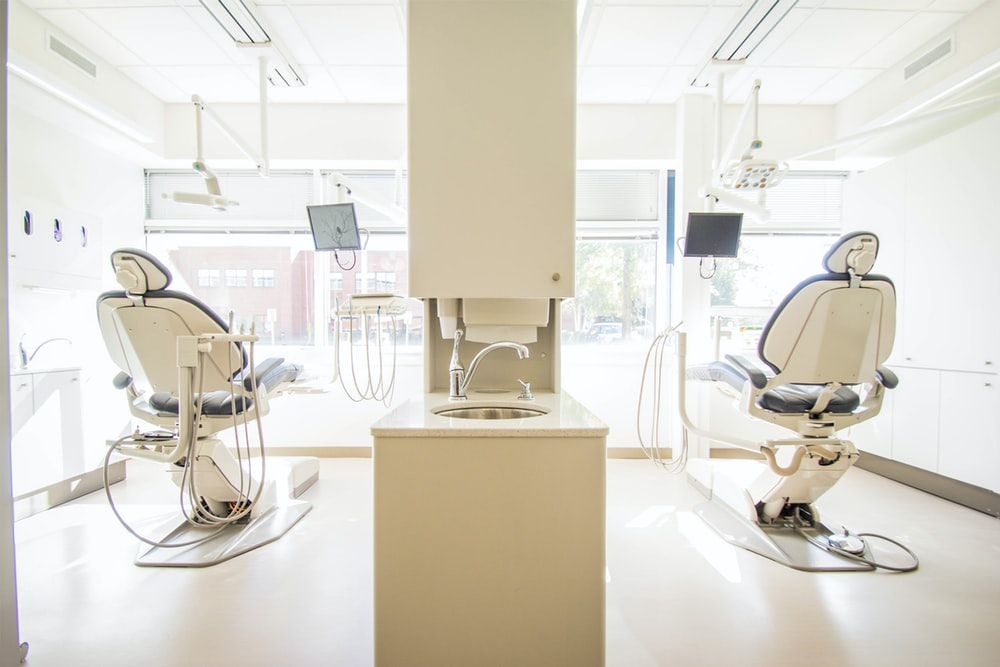
What Are Digital Dental X-Rays?
Digital dental x-rays, also called digital radiographs, utilize electronic sensors instead of photographic film to capture images. The main types used in dentistry include:
- Intraoral Sensors: These are placed inside the mouth and are primarily used to capture images of teeth, bone, and soft tissues. They provide detailed views that are crucial for accurate diagnosis and treatment planning.
- Extraoral Sensors: These are used externally to scan larger areas such as the skull and jawbones. They offer a comprehensive view of the patient's oral structure.
- Phosphor Storage Plates: These are reusable plates that temporarily store latent images before they're processed digitally.
A distinct feature of digital sensors is their ability to directly convert x-rays into digital data. This data can then be viewed on a computer, allowing easy storage and retrieval. This is a significant advancement from traditional film x-rays, which require chemical processing to develop the images.
Benefits Of Digital Dental X-Rays
Digital radiography offers many benefits that significantly enhance the efficiency and effectiveness of dental care.
- Reduced Radiation Exposure: Digital sensors used in digital dental x-rays are more sensitive than traditional film, using significantly less radiation per exposure. This safely minimizes the patient's exposure to radiation.
- Instant Viewing And Patient Education: Images captured by digital sensors are displayed instantly on a computer screen, eliminating the wait time associated with film processing. This real-time image display, along with tools like intraoral cameras, aids in patient consultations, allowing my general dentist in Thornhill to explain diagnoses and treatment plans more effectively.
- Enhanced Images: Software filters enhance the contrast and clarity of the images. Features such as zoom and colorization further optimize the view, providing superior image quality for more accurate diagnoses.
- Space Savings And Cost Savings: Digital x-rays are stored digitally, which not only saves physical space by eliminating bulky film files but also integrates easily into patients' electronic records. Over time, digital systems can lead to cost savings by avoiding expenses related to film, chemicals, and storage.
- Simplified Sharing: The digital nature of these x-rays allows for easy integration into electronic patient records and instant sharing with specialists when needed.
- Reduced Waste: Digital x-rays are environmentally friendly. They avoid the chemical waste associated with film processing and reduce the need for retakes, contributing to sustainability in dental practices.
These advancements have enhanced the quality of dental imaging while contributing to patient safety, education, and overall experience. The use of digital dental x-rays highlights the ongoing progress in dental technology.
Considerations When Switching To Digital
While the benefits of digital dental x-rays are numerous, transitioning from traditional film to digital radiography comes with its own set of challenges. It's a process that requires careful consideration of several factors:
- Equipment Investments And IT Requirements: Transitioning to digital radiography involves significant upfront costs for digital sensors, software, computers, and accessories. Additionally, appropriate IT infrastructure and cybersecurity measures are essential to protect patient data.
- Learning Curve: Staff will need training in image acquisition, usage of features, and digital workflow.
- Storage Needs: With digital images, server and cloud storage options must be considered to provide adequate long-term capacity.
- Malpractice Insurance: It's important to confirm that your coverage includes digital radiography and records storage.
- Portability Limitations: Wired sensors offer limited mobility compared to traditional film packets, although wireless options are available.
These challenges notwithstanding, careful planning and consideration can help ensure a smooth transition. The benefits of improved image quality, reduced radiation exposure, and enhanced patient communication make this investment worthwhile for the future of dental care.
Positioning Sensors For Optimal Images
To capture optimal images using digital dental x-rays, the correct positioning of the sensors is crucial. It minimizes the need for retakes, thus reducing radiation exposure. Here are important guidelines to follow:
- Select a properly sized sensor for the area of interest.
- Fully seat sensors against teeth with gentle pressure.
- Ensure sensors lie parallel to the tooth plane.
- Use proper perpendicular beam alignment without angulation.
- Employ holding devices as needed to stabilize sensors.
- Follow any special techniques for incisors, bitewings or edentulous (missing all teeth) patients.
With careful attention to detail and adherence to prescribed techniques, dental professionals can capture high-quality images that aid in accurate diagnosis and effective treatment planning.
Digital Radiography Best Practices
To maximize the benefits of digital radiography, consider the following best practices:
- Train Staff: Ensure all staff are trained in ideal scanning and image handling protocols.
- Post Instructions: Display detailed imaging instructions visibly in operatories for constant guidance.
- Evaluate Quality: Regularly assess the diagnostic quality of the x-rays and retake any that are inadequate.
- Leverage Tools: Utilize the built-in image enhancement and viewing tools to optimize the images for better diagnosis.
- Develop Workflows: Establish standardized workflows for efficient image management.
- Backup Data: Securely store images on both local servers and cloud storage to ensure data safety and accessibility.
- Use Software: Implement integrated imaging software for seamless sharing and annotation to enhance communication.
These best practices can significantly improve the efficiency and effectiveness of digital radiography in dental care.
Final Thoughts
Transitioning to digital radiography is a strategic investment that brings significant benefits for both dentists and patients. By adhering to proper techniques and workflows, digital systems can greatly enhance dental care outcomes.
Despite the initial learning curve, this advancement promises a future of improved diagnosis and treatment planning, making it a positive step forward in dentistry.
Author Bio: Michael Miller is a seasoned content writer with a keen interest in healthcare and technology. Outside of writing, Michael enjoys staying updated on the latest tech trends, enriching both his professional insights and personal interests.