A new study has just been published in the journal NeuroImage that strongly suggests magnetoencephalography (MEG for short) can be used as a therapeutic tool to control and train targeted brain regions. The study was written by and based on research from the Montreal Neurological Institute and Hospital, McGill University and the McGill University Health Center. The research and report are the first to clearly demonstrate that a new and advanced brain imaging technique based on MEG - that enables people to view their brain activity in real time and that provides the ability to both control and adjust function in pre-determined brain regions - has important clinical applications for numerous neurological and neuropsychiatric conditions.
MEG is a non-invasive imaging technology that measures magnetic fields generated by nerve cell circuits in the brain. MEG captures these tiny magnetic fields with noteworthy accuracy and amazing time resolution - a millisecond time scale across the entire brain.
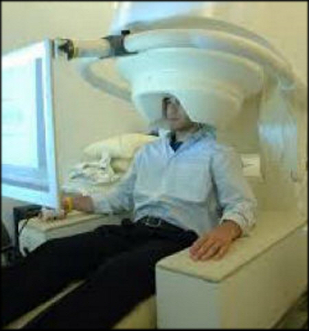
The research, which serves essentially as a proof of concept, participants had nine sessions in "the MEG" (see the image below) and used neurofeedback to reach a specific target. The target consisted of having the participants look at a colored disc on a display screen and having them find their own strategies to change the disc's color from dark red to bright yellow white, and to maintain that bright color for as long as possible.
Dr. Sylvain Baillet, acting Director of the Brain Imaging Centre at Montreal Neurological Institute and Hospital and the lead investigator on the study, notes that, "With MEG participants or patients can observe their own brain activity as it happens in real time. We can use MEG for neurofeedback. This is a process by patients can see on-going physiological information that they aren't usually aware of - in this case their own brain activity - and use that information to train themselves to self-regulate.
The disc color in the proof of concept experiments was indexed on a very specific aspect of ongoing brain activity from the participants. The researchers had set it up so that the experiment would access predefined regions of the motor cortex in each participant's brain. The color presented changed according to a predefined combination of slow and faster brain activity within these regions. This was possible because the researchers combined MEG with MRI, which provides information on the brain's structures, known as magnetic source imaging (MSI). The research findings pave the way to use MEG as an innovative therapeutic approach for treating patients.
Dr. Baillet notes that for him the remarkable thing is that with each training session the participants were able to reach the target aim faster, even though the researchers kept raising the bar for the target objective in each session. These results demonstrate that participants were successfully using neurofeedback to alter their pattern of brain activity according to a predefined objective in specific regions of their brain's motor cortex, without moving any body part. More specifically, this demonstrates that MEG source imaging can provide brain region-specific real time neurofeedback and that longitudinal neurofeedback training is possible with this technique.
Many Therapies Possible
To date, work with epilepsy patients has shown the most promise but there is no reason to believe it won't also work with other neurological syndromes and neuropsychiatric disorders that include stroke, dementia, movement disorders and chronic depression - to name but a few. MEG has the potential to reveal dynamics of brain activity involved in perception, cognition and behavior and has provided unique insight on both brain functions such as language, motor control, visual and auditory perception, as well as brain dysfunctions - such as movement disorders, tinnitus, chronic pain and dementia.
There are other related applications. For example, MEG looks promising as a therapy for people that have amusia, a disorder that makes them unable to process musical pitch. Amusia is possibly due to fpoor connectivity between the auditory cortex and prefrontal regions in the brain. Working with Prof. Isabelle Peretz at Université de Montréal, Dr. Baillet and the research team are using MEG to measure the intensity of functional connectivity between these brain regions in amusic patients and aged-matched healthy controls.
Using MEG and neurofeedbackthe team hopes to take advantage of the brain's "plasticity" to reinforce the functional connectivity between the target brain regions. If the approach demonstrates an improvement in pitch discrimination in participants, that will demonstrate the clinical and rehabilitative applications of this approach. The baseline measurements have been taken already, and the training sessions will take place over this year.
There is a great deal of work yet to do but the results from the current experiments are nothing short of hugely encouraging. Dr. Baillet underscores that, "Our ultimate hope and aim is to enable patients to train specific regions of their own brain, specifically in a way that relates to their particular condition." Amen to that - we certainly wish Dr. Baillet Godspeed in turning the research into real world therapies.
Edited by
Cassandra Tucker