Recently we wrote about advanced new brain imaging technology that provides amazing views into how the brain is wired, and how signals travel along those “wires” as synapses fire, send signals out and make connections. It represents a collection of truly remarkable achievements.
Now we’ve got yet another amazing technology breakthrough on our hands – this one focused on actually making parts of the brain transparent. That’s right, we mean the brain’s tissues become see-through, allowing scientists and doctors to actually view and analyze the brain in entirely new ways.
The new technology, dubbed Clarity by its Stanford University inventors, was recently elaborated upon in a detailed article in the journal Nature. Making the brain tissue transparent doesn’t mean it makes it invisible – rather it means that the internal connections within the brain can actually be viewed in full 3D, and further they can be viewed in full color. It is quite a leap forward for studying the brain; with the technology there is no longer a need to physically slice up cross sections to be viewed under microscopes.
The Stanford scientists have been able to render an entire mouse brain transparent, as well as a part of a human brain.
Back in the old days, we once assembled a see-through and fully working model of a V8 car engine. The new brain technology is uncannily similar to this – following the very short video below, keep it in mind when reading the rest of the article.
Just for the fun of it, here’s the actual Nature summary of the article:
Obtaining high-resolution information from a complex system, while maintaining the global perspective needed to understand system function, represents a key challenge in biology. Here we address this challenge with a method (termed CLARITY) for the transformation of intact tissue into a nanoporous hydrogel-hybridized form (crosslinked to a three-dimensional network of hydrophilic polymers) that is fully assembled but optically transparent and macromolecule-permeable.
Using mouse brains, we show intact-tissue imaging of long-range projections, local circuit wiring, cellular relationships, subcellular structures, protein complexes, nucleic acids and neurotransmitters. CLARITY also enables intact-tissue in situ hybridization, immunohistochemistry with multiple rounds of staining and de-staining in non-sectioned tissue, and antibody labeling throughout the intact adult mouse brain.
Finally, we show that CLARITY enables fine structural analysis of clinical samples, including non-sectioned human tissue from a neuropsychiatric-disease setting, establishing a path for the transmutation of human tissue into a stable, intact and accessible form suitable for probing structural and molecular underpinnings of physiological function and disease.
The bolding of the words above is our own, and highlights the key aspects of what the new technology is able to accomplish – these are fairly self-explanatory. The words we did not highlight more or less mean that the new technology essentially manages to safeguard the brain’s own original biochemistry. That is, the transparent model remains quite robust and will stand up to numerous tests.
For example, doctors and researchers can view the results of applying certain chemicals that highlight specific structures within the model. What’s more, the new technology can be applied to brains that have long been stored (yes, scientists preserve and store brains).
This allows scientists to both learn entirely new things about the way the normal brain works, and allows them to also compare this newly visualized “normal baseline” to how non-normal brains operate or are built or otherwise function. The process will hopefully begin to uncover the myriad of underlying problems and mysteries associated with, for example, mental disorders.
What’s different about the brains of people diagnosed as being bi-polar, or schizophrenic? The ability to uncover real answers to these and numerous other brain-related disorders – as well as the perhaps even more critical ability to document how healthy brains work – has taken a huge leap forward.
It has taken years to develop the process, which requires a variety of steps that include the proper application of an electrical current within a proper temperature range. That certainly does sound a lot like Frankenstein, we know. Once again, science fiction turns into reality.
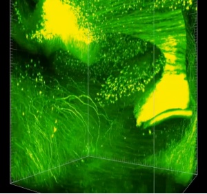
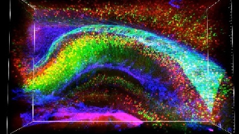
Coupled with the work being done to create the brain’s wiring schematics noted in the earlier article mentioned above, scientists and researchers look to be well on their way to opening significant new doors to understanding the brain, understanding the normal brain and truly beginning to figure out what can go wrong, what does go wrong – and most importantly, how to put things back in order. Below are shown two admittedly poor quality screen captures from a video and images that are available at Nature.
We won’t try to describe them, so check out the videos and images themselves for details.
The Nature Clarity article – including detailed photos and videos – is available for purchase for a modest price and is well worth reading.
Edited by
Braden Becker